Showing 1–12 of 13 resultsSorted by latest
-
AED 714.00
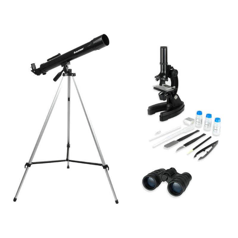
- This 3-in-1 kit contains everything young scientists need to explore the world around them
- 50mm refractor telescope perfect for viewing terrestrial and celestial objects
- Microscope offers up to 900x magnification and accessory kit
- 4×30 binoculars
-
AED 693.87
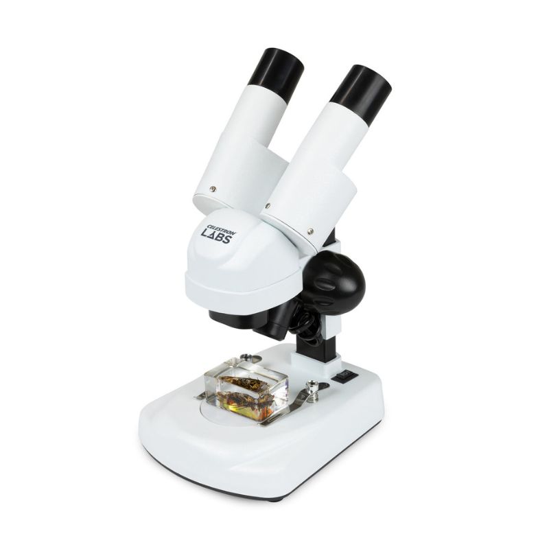
- Stereo microscope with 20x power
- Ergonomic angled binocular viewing head
- 10x eyepieces with 2x objective lenses
- Greenough objective design with upper and lower LED illumination
- Powered by 2AA batteries for portability
- Accessories include two 3D insect specimens, two stage plates, and a dust cover
-
Sale!
AED 484.84 Original price was: AED 484.84.AED 381.02Current price is: AED 381.02.
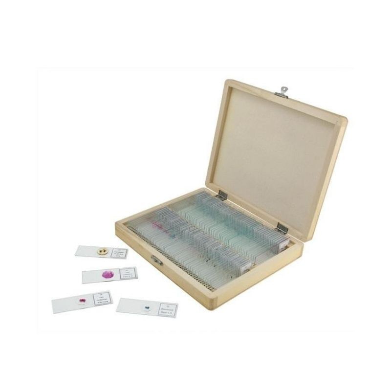
- Prepared Slides For Hours and Hours of Enjoyment and Discovery
- 1″ x 3″ (25 mm x 76 mm) Glass Slides
- Packed in a Wooden Case
- Listing of each of the Slides
-
AED 136.00
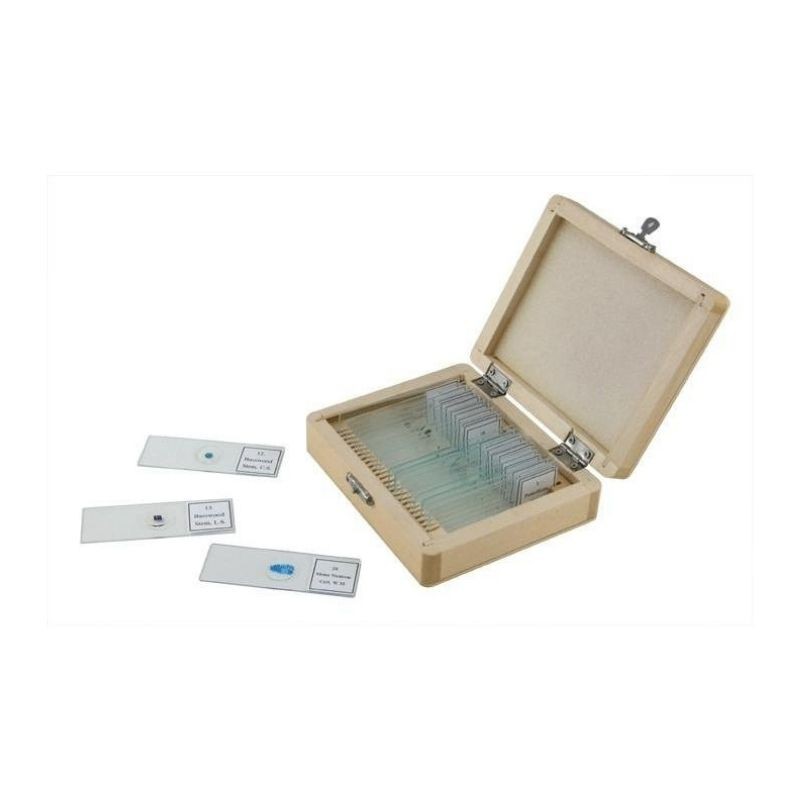
- Prepared Slides For Hours and Hours of Enjoyment and Discovery
- 1″ x 3″ (25mm x 76mm) Glass Slides
- Packed in a Wooden Case
- Listing of each of the Slides
-
AED 366.09
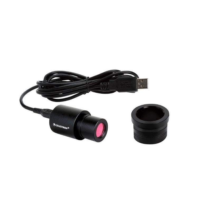
- 2MP image sensor makes a traditional microscope into a digital microscope.
- Capture images and video of your specimens via your computer.
- The microscope imager works with 23 mm to 30 mm diameter eyepieces.
- Connect the imager to your computer via USB 2.0 cable.
- Downloadable software works with both Mac and Windows PCs
- Imager Works with Mac OS via PhotoBooth and Quick Camera.
-
AED 590.27
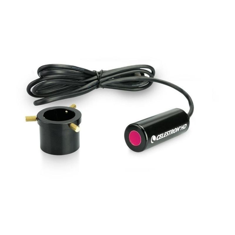
- Simple, rugged aluminum housed 5MP CMOS sensor
- Replaces the eyepiece on your traditional microscope. Turns any existing traditional microscope with 23mm or 30mm eyepiece diameters into a digital microscope
- Takes high resolution still images and 30 fps video*
- USB powered – Connects to your personal computer via open USB port
- Downloadable software works with both Mac and Windows PCs
- More robust software with new features like measurement, calibration, note taking and comparison feature which allows comparing 2 live streams
-
Sale!
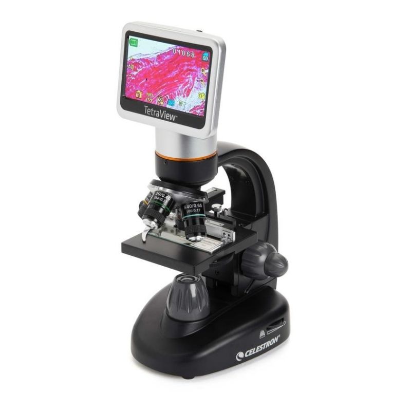
AED 2,433.36 Original price was: AED 2,433.36.AED 1,999.00Current price is: AED 1,999.00.
- 40x to 600x magnification, up to 2400x with digital zoom
- 4.3″ LCD touchscreen
- 5MP CMOS built-in digital camera
- 5 fully achromatic objective lenses: 4x, 10x, 20x, 40x, 60x
- Top & bottom adjustable LED illumination
- 180° rotatable LCD screen
- Fully mechanical stage
- Coarse and fine focus knobs
- Filter wheel/diaphragm
- AV/TV output with cable
- Optional battery mode
- SD card slot with 4GB SD card included (Supports up to 32GB)
- Universal AC adapter with multi-plugs for international usage
- Accessories included: 10 prepared slides, hard carrying case, dust cover, touch pen, USB cable, AV cable
-
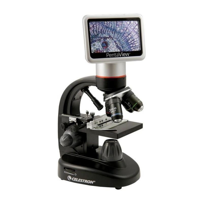
AED 2,084.00
- 40x to 400x magnification (up to 1600x with digital zoom)
- 4.3″ LCD touchscreen
- 5MP CMOS built-in digital camera
- 4 fully achromatic objective lenses: 4x, 10x, 20x, 40x
- Adjustable LED bottom illumination
- 180° rotatable LCD screen
- Fully mechanical stage
- Coarse and fine focus knobs
- Filter wheel/diaphragm
- AV/TV output
- Battery power option (4AA batteries)
- 2GB SD card included (SD card slot supports up to 32GB)
- Universal AC adapter with multi-plugs for international usage
- Accessories included: 7 prepared slides, hard carrying case, dust cover, touch pen, USB cable, AV out cable
-
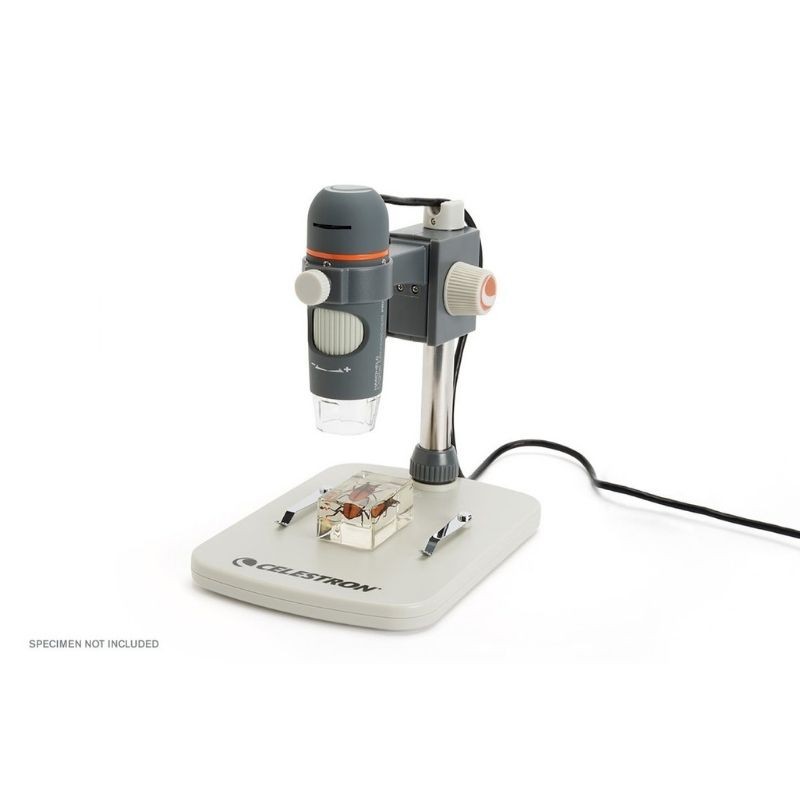
AED 1,199.00
- 1080p video streaming of microscopic worlds via included HDMI cable directly to your monitor or projector. NO COMPUTER NEEDED!
- 3.5MP high speed sensor has larger pixel sizes that allow more details and a higher refresh rate
- 10x to 220x power magnification
- Records high definition still images. Images are saved directly to a Micro SD card (not included)
- Capture 720p High Definition video at 30 fps when connected to a PC via USB using the downloadable Celestron Portable Capture HD software.
-
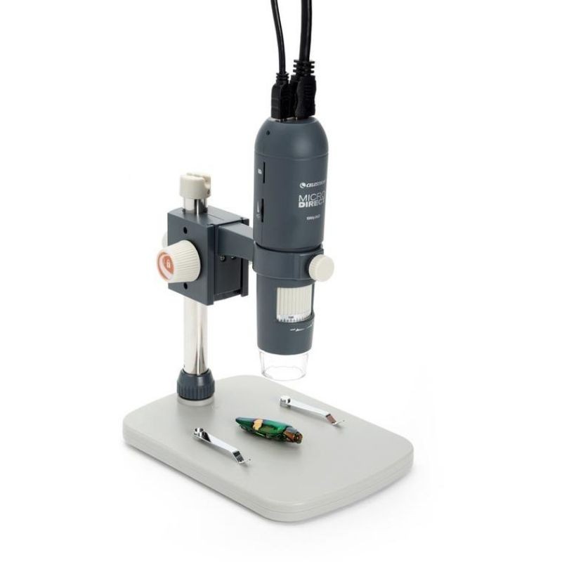
AED 749.00
- True 5MP sensor to capture and save high-resolution images and videos of your specimens
- 5-Element IR cut high-quality glass lens ensures sharper images
- 20x to 200x powers, great for low-power observation of 3D specimens (Note: Final magnification determined by monitor size)
- Professional, adjustable height stand included for hands-free operation
- 4 foot USB 2.0 cable for easy maneuverability when viewing large surfaces
- Downloadable MicroCapture Pro software available for both Mac and Windows PCs
-
Sale!
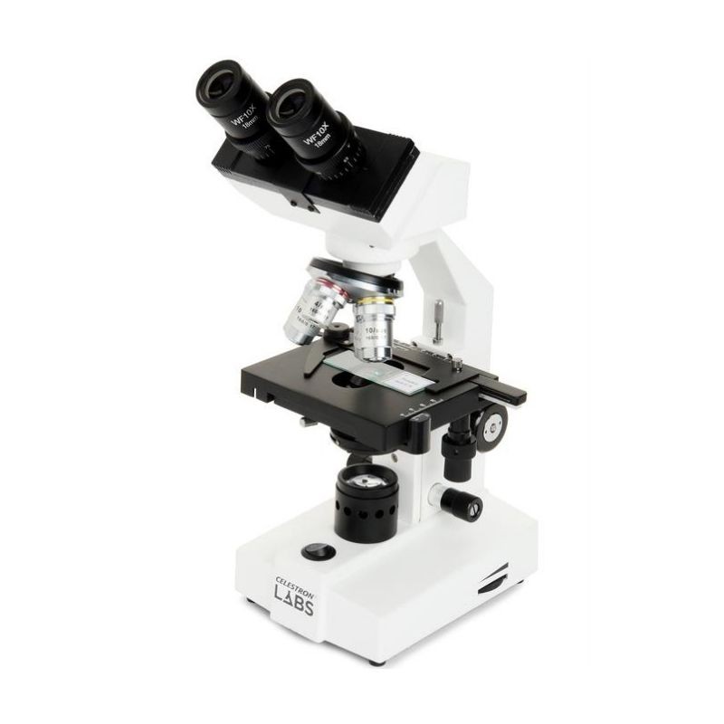
AED 1,966.23 Original price was: AED 1,966.23.AED 1,899.00Current price is: AED 1,899.00.
- Lab-ready compound binocular microscope with up to 1000x power
- 4 fully achromatic objective lenses- 4x, 10x, 40x, 100x
- Coarse and fine focus knobs
- 10x eyepieces allow you to view specimens at 40x, 100x, , 400x, , and 1000x magnification
- Adjustable binocular head
- All-metal construction with fully mechanical stage
- Built-in, adjustable LED lower illumination with Abbe condenser and iris diaphragm
-
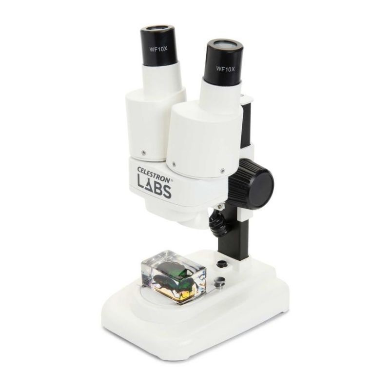
AED 497.18
- Great introductory stereo microscope with 20x power
- 10x eyepieces with 2x objective lenses
- Metal head with coarse focusing knob
- Upper LED illumination for detailed viewing
- Two insect specimens included
- Two AA batteries included